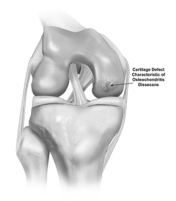
Osteochondritis Dissecans (OCD)
There are three bones that make up the knee joint – the femur (thigh bone), the tibia (shin bone), and the patella (kneecap). Articular cartilage covers both the bones and functions to reduce friction for smooth joint movement.
Osteochondritis dissecans (OCD) is a joint disorder in which a fragment of cartilage and underlying bone is detached from the joint surface. OCD occurs when a small segment of bone and cartilage, usually at the end of a bone, loses its blood supply. Without adequate blood flow, the affected bone and cartilage can deteriorate and detach from the underlying bone.

The exact cause of osteochondritis dissecans is not always clear, but it is believed to involve a combination of genetic, mechanical, and vascular factors. OCD is more common in adolescents and young adults, especially those involved in sports that place repetitive stress on the affected joint.
- Joint Pain: OCD typically causes pain in the knee joint. The pain is typically a dull ache, with intermittent sharp pain, felt on either side of the knee or in the front of the knee. The pain often worsens with activity and improves with rest.
- Stiffness: People with OCD may experience stiffness in the knee joint, making it difficult to move the knee through its full range of motion.
- Reduced Range of Motion: As the condition progresses, the range of motion in the knee joint may become limited, making it challenging to perform activities like walking, bending, or climbing stairs.
- Swelling.
- Catching/locking: Catching or locking of the knee joint can occur in cases where the OCD piece or fragments of the OCD gets dislodged is present within the knee joint.
Physical Exam
A comprehensive physical exam will be conducted to assess the range of motion in your knee joint, look for signs of inflammation or swelling, and feel for areas of tenderness or crepitus (grating or grinding sensations). Your gait (the way you walk) will also be evaluated to identify any abnormalities.
Imaging
Diagnostic imaging is necessary to definitively diagnose OCD of the knee. X-rays will allow Dr. Chahla to assess the degree of remaining joint space, the extent of cartilage damage, and subchondral involvement. Dr. Chahla will also recommend an MRI of the knee to further assess the size and extent of the OCD lesion, as well as subchondral involvement and any possible concomitant injuries to the meniscus or surrounding structures.
Osteochondritis dissecans (OCD) is a condition where a piece of cartilage and underlying bone in the knee or hip joint becomes damaged due to lack of blood supply, leading to pain, swelling, and joint instability. If left untreated, OCD can result in loose bone fragments and long-term joint damage. Dr. Jorge Chahla, an expert orthopedic knee and hip surgeon, provides advanced treatment options to restore joint health. If you are experiencing joint pain, swelling, or locking, schedule an appointment with Dr. Chahla in Chicago, Naperville, or Oak Brook for an evaluation.
At a Glance
Dr. Jorge Chahla
- Triple fellowship-trained sports medicine surgeon
- Performs over 700 surgeries per year
- Associate professor of orthopedic surgery at Rush University
- Learn more

