Avascular Necrosis (AVN)
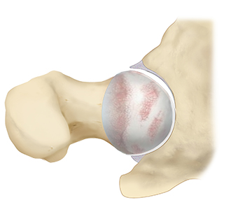
The hip is a ball and socket joint. The socket is the part of the hip bone called the acetabulum, and the head of the femur is the ball. Articular cartilage, which is a shiny white tissue, covers both the ball and the socket and functions to reduce friction for smooth joint movement. The health of the bone beneath the articular cartilage is important to maintain a smooth, congruent joint surface.
Avascular necrosis, or osteonecrosis, of the hip occurs when the blood supply to the femoral head is disrupted, leading to a lack of nutrients and oxygen delivery to the bone. Without these necessities, the bone dies. Over time, the dead bone weakens and eventually collapses, interfering with the articular cartilage on the surface. This leads to progressive osteoarthritis and destruction of the hip which can be painful and disabling.
AVN of the femoral head is a debilitating, progressive condition that occurs in up to 20,000 people in the United States per year.

AVN can occur in anyone, but it is most common in individuals between the ages of 40 and 65. It is also more common in men than women. In many instances, both hips may be affected by AVN.
There have been several risk factors that have been identified for hip AVN. Most commonly, patients who develop AVN have a history of excessive alcohol use or chronic corticosteroid use. Both substances lead to fatty deposits in the bone marrow which result in decreased blood flow to the bone. Trauma to the hip, such as a dislocation or fracture, is another known cause of AVN. Trauma can damage the blood vessels in the femoral head and disrupt its blood supply. Although rarer, a handful of medical conditions have also been associated with the development of AVN. These include Caisson disease (“the bends”), sickle cell disease, myeloproliferative disorders, Gaucher’s disease, systemic lupus erythematosus, Crohn’s disease, arterial embolism, thrombosis, and vasculitis.
AVN usually presents initially with new-onset hip pain located in the groin. This may progress to dull aching or throbbing pain in the groin. As hip AVN progresses, it is more difficult to walk or bear weight on the affected hip.
It may take anywhere from a few months to a year for the disease to progress. Early diagnosis is important because some studies have shown better outcomes when treated earlier in the AVN disease process.
During your consultation with Dr. Jorge Chahla, a Chicago sports medicine surgeon and AVN specialist, your medical history, AVN symptoms, and diagnostic imaging will be reviewed.
The diagnosis of AVN relies heavily on imaging studies. An x-ray of the hip is the initial study to examine the bony structures of the joint. This allows Dr. Jorge Chahla to determine if the bone in the femoral head has collapsed and to what degree. A common x-ray finding is a wedge-shaped area of dense, whitish substance in the superior lateral portion of the femoral head.
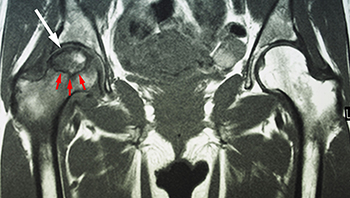
However, if the x-ray is normal, an MRI of the hip is obtained to look for early signs of AVN. An MRI has the added advantage of picking up signs of AVN that would be otherwise missed on x-ray. This is especially helpful in discovering lesions of AVN that have not yet developed symptoms, as in a lesion on the opposite hip. MRI may also be used to determine how much of the bone is affected.

Avascular necrosis (AVN) occurs when reduced blood flow to the hip joint leads to bone death and joint deterioration. This condition can cause progressive hip pain, stiffness, and loss of function. Dr. Jorge Chahla is a leading orthopedic hip specialist who provides advanced diagnostic and treatment options for AVN to help prevent further joint damage. If you are experiencing persistent hip pain, schedule an evaluation with Dr. Chahla in Chicago, Naperville, or Oak Brook to discuss the best course of action.
At a Glance
Dr. Jorge Chahla
- Triple fellowship-trained sports medicine surgeon
- Performs over 800 surgeries per year
- Associate professor of orthopedic surgery at Rush University
- Learn more


