Failed Hip Arthroscopy
What is FAI?
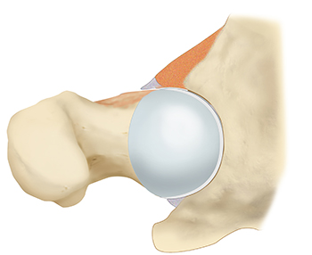
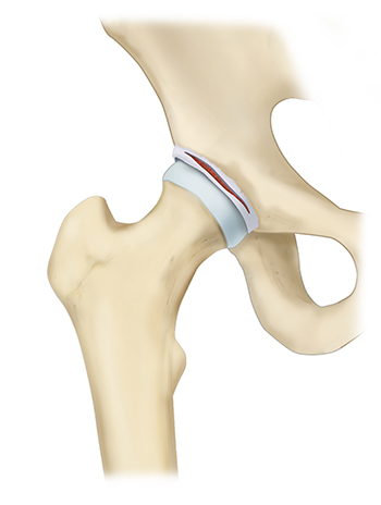
FAI (Femoroacetabular Impingement) is a common hip joint disorder in young, active patients caused by a structural problem, specifically abnormal bone growth, that leads to irregular contact between the acetabulum and the femoral head. The hip is a ball and socket joint. The socket is the part of the hip bone called the acetabulum, and the head of the femur is the ball. Articular cartilage covers both the ball and the socket and functions to reduce friction for smooth joint movement. In a healthy hip, the ball of the femur fits perfectly into the acetabulum. However, in individuals with FAI, there may be irregularities in the shape of either the ball or the socket, or both. These irregularities cause hip joint impingement during activities like walking, running, or bending the hip, which can result in hip impingement symptoms such as groin pain and discomfort.
The location of the hip impingement can be described as either CAM impingement or Pincer impingement, or both. The CAM type is caused by the abnormal shape of the femoral head and neck, while the Pincer-type is caused an abnormal shape of the acetabulum. In some cases, both types can co-exist.

The friction and pressure caused by FAI can lead to further hip joint damage over time, including bone spurs, a labral tear, and hip osteoarthritis. Thus, early hip impingement diagnosis by a hip impingement specialist is crucial.
The hip labrum is a ring of cartilage that surrounds the hip socket, providing stability to the joint. The hip labrum plays an important role in maintaining normal hip function. It functions to tighten the seal between the bones for joint stability, allows for a wide range of motion, and helps to maintain the alignment between the bones. A hip labral tear occurs when this cartilage is damaged or torn, often due to a hip injury, degeneration, or FAI. This tear can lead to a range of symptoms, including hip labrum pain (groin pain), clicking or catching sensations, and a decreased range of motion.

The hip capsule is a crucial anatomical structure that provides stability, protection, and lubrication to the hip joint, enabling smooth and controlled movement of the leg while also preventing excessive motion and potential injuries. The hip capsule, also known as the acetabular capsule, is a fibrous, ligamentous structure that surrounds and stabilizes the hip joint. It consists of strong connective tissue and ligaments that form a capsule-like structure around the ball-and-socket joint of the hip. The functions of the hip capsule include:
Joint Stability
The hip capsule plays a crucial role in maintaining the stability of the hip joint. It helps keep the head of the femur securely within the socket of the acetabulum. This stability is important for weight-bearing, movement, and activities such as walking, running, and jumping.
Lubrication and Nutrition
The synovial membrane lining the inside of the hip capsule produces synovial fluid. This fluid lubricates the joint, reducing friction between the articulating surfaces and ensuring smooth movement. It also provides essential nutrients to the cartilage within the joint, helping to maintain its health.
Joint Protection
The capsule acts as a protective barrier for the hip joint, helping to shield it from external forces and injuries.
Limiting Excessive Movement
While allowing for a wide range of motion, the hip capsule also limits excessive movement of the joint, preventing dislocations and maintaining joint integrity.
Proprioception
The ligaments within the hip capsule contain sensory receptors known as proprioceptors. These receptors provide feedback to the nervous system about the position and movement of the hip joint. This information helps with balance, coordination, and posture.
Re-injury to the hip labrum or hip capsule following hip arthroscopy can occur for a variety of reasons. Most commonly, patients may recall a trauma or injury in which the operative leg was forced into either an extreme degree of extension or external rotation. Additionally, failed hip arthroscopy can occur due to the presence of pre-existing risk factors such as hyper-laxity (Ehlers Danlos syndrome), untreated hip dysplasia, osteoarthritis, or a degenerative labrum.
Symptoms of a failed hip arthroscopy include:
- Intermittent deep groin pain or ache (most common)
- Pain at the outside of the hip joint
- Sharp stabbing pain when twisting, turning, or squatting, such as when getting in or out of a car or a chair.
- A dull ache from prolonged sitting or walking
- A sensation of catching, clicking, or locking in the hip joint during movement
- Instability of the hip joint
- Stiffness and reduced flexibility in the hip joint
- Limping
During your consultation with hip surgeon Dr. Jorge Chahla your medical and surgical history, FAI symptoms, and past injuries will be reviewed. It is highly recommended to bring all records from your previous FAI surgery, including the operative report, any surgical photos, and any subsequent imaging, to your consultation with Dr. Chahla, as this information can be very helpful in the diagnostic process.
Physical Exam
He will conduct a physical examination checking range of motion including flexion, adduction, and rotation to diagnose your hip impingement. The hip impingement test will also be performed during your physical exam. This is a test in which Dr. Chahla will ask you to bring your knee to your chest while Dr. Chahla rotates the knee in toward the opposite shoulder (FADIR [flexion, adduction, and internal rotation]). If the hip impingement test causes pain, you likely have a hip labral tear. A comprehensive physical exam is key to determine the cause of your hip pain.
Imaging
Diagnostic imaging is necessary to definitively diagnose FAI and a hip labral tear. X-rays will reveal abnormally shaped bones, and an MRI will reveal damage to the soft tissue, such as a re-tear of the labrum or an injury to the hip capsule. A CT scan may be necessary to further evaluate details about the shape of the bones, location of abnormal bone growth, and the rotation of the bones that may have contributed to the re-injury.
Hip Injection
An intra-articular hip injection can help to confirm the diagnosis of a failed hip arthroscopy. This injection will be performed in the office at the time of your office visit under ultrasound guidance. If the majority of your pain goes away, even temporarily, following the injection, then it confirms that the source of the pain is due to a possible re-tear of the labrum or capsular injury.
A failed hip arthroscopy occurs when previous hip surgery does not fully resolve symptoms or leads to persistent pain and dysfunction. This can be due to incomplete correction of structural issues, residual impingement, or post-surgical complications. Hip surgeon Dr. Jorge Chahla specializes in diagnosing and treating patients who have experienced failed hip arthroscopy, offering revision surgery and alternative treatment options to restore hip function. If you are still struggling with hip pain after surgery, contact hip surgeon Dr. Chahla’s office in Chicago, Naperville, or Oak Brook for expert evaluation and treatment.
At a Glance
Dr. Jorge Chahla
- Triple fellowship-trained sports medicine surgeon
- Performs over 800 surgeries per year
- Associate professor of orthopedic surgery at Rush University
- Learn more


