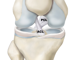
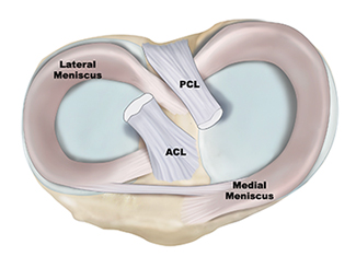
ACL Anatomy
There are three bones that make up the knee joint – the femur (thigh bone), the tibia (shin bone), and the patella (kneecap). There are two cruciate ligaments—Anterior Cruciate Ligament (ACL) and Posterior Cruciate Ligament (PCL)—inside the knee joint that cross to form an X. The ACL sits in the front of the knee joint, while the PCL sits in the back of the knee joint. Together, they help control the front-to-back motion of the knee, as well as rotation.
Schedule a knee consultation
If you or a family member have suffered an ACL injury, schedule a consultation with Dr. Jorge Chahla. Dr. Jorge Chahla is a highly regarded knee surgeon with offices in Chicago, Naperville, and Oak Brook. He focuses on the comprehensive diagnosis and advanced treatment of ACL injuries, helping patients regain stability and function. If you are experiencing knee instability, swelling, or challenges with weight-bearing, schedule a consultation with Dr. Chahla to explore personalized treatment options and begin your path toward restoring knee health.
- Triple fellowship-trained sports medicine surgeon
- Performs over 800 surgeries per year
- Associate professor of orthopedic surgery at Rush University
- Learn more


