Pediatric ACL Injuries
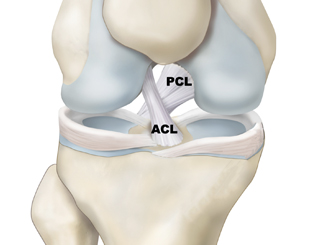
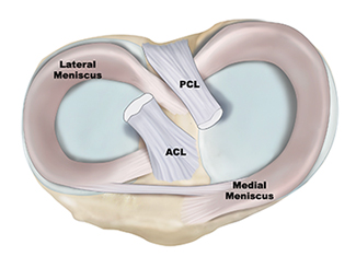
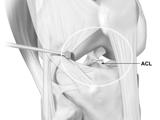
There are three bones that make up the knee joint – the femur (thigh bone), the tibia (shin bone), and the patella (kneecap). There are two cruciate ligaments—Anterior Cruciate Ligament (ACL) and Posterior Cruciate Ligament (PCL)—inside the knee joint that cross to form an X. The ACL sits in the front of the knee joint, while the PCL sits in the back of the knee joint. Together, they help control the front-to-back motion of the knee, as well as rotation.
ACL injuries can be classified into different types based on the severity and extent of the damage to the ACL fibers. The grading of severity is based on the amount of ligament disruption and the extent of knee instability present following an injury. It is important to accurately diagnose the extent of an ACL injury, as the appropriate treatment plan can vary based off the type of ACL injury that an individual has suffered.
Grade 1: ACL Sprain
An ACL injury that stretches the fibers of the ligament but does not lead to a full thickness tear of the fibers is called an ACL Sprain. This is the least severe injury to the ACL, and often, these injuries can heal on their own with conservative treatment including rest, ice, compression, elevation, oral anti-inflammatory medications, and physical therapy.
Grade 2: Partial ACL Tear
An ACL injury that tears some of the ligament fibers is called a “partial tear” of the ACL. When partial tearing of the ACL occurs, patients may experience some episodes of mild instability or pain. However, the severity can vary depending on the extent of the partial tear. In some cases, a partial tear of the ACL may heal with conservative treatment including rest, ice, compression, elevation, oral anti-inflammatory medications, physical therapy, and possibly the need of a specialty brace called a functional ACL brace. However, when instability persists despite conservative treatment, it is reasonable to consider surgical intervention. The decision to perform surgery to fix a partial tear depends on the severity of instability of the knee and patient’s desire to return to sport. When discussing surgery for a partial ACL tear, knee surgeon Dr. Chahla and his team may discuss both the option of an ACL repair and an ACL reconstruction. The options available to you for surgery will depend on the location and extent of your partial ACL tear. Please see treatment options below for further information.
Grade 3: Complete ACL Tear
A complete tear of the ACL describes an injury in which the ligament is non-functional and will require surgery to fix it. Professional athletes and most recreational athletes will likely require surgery to return to play, especially if their desired sport includes pivoting, cutting, and lateral movements.
Children and adolescents are increasingly at risk for ACL injuries due to the rise in competitive sports and high-impact activities. These injuries require specialized treatment to protect knee function and prevent future joint problems. Dr. Jorge Chahla is a board-certified orthopedic knee surgeon that is experienced in treating pediatric ACL injuries, offering both surgical and non-surgical approaches tailored to growing athletes. If your child has suffered a knee injury and needs expert care, schedule an appointment with Dr. Chahla at his offices in Chicago, Naperville, or Oak Brook to ensure the best possible treatment.
- Triple fellowship-trained sports medicine surgeon
- Performs over 800 surgeries per year
- Associate professor of orthopedic surgery at Rush University
- Learn more


