Quantitative and Qualitative Analysis of the Medial Patellar Ligaments: An Anatomic and Radiographic Study
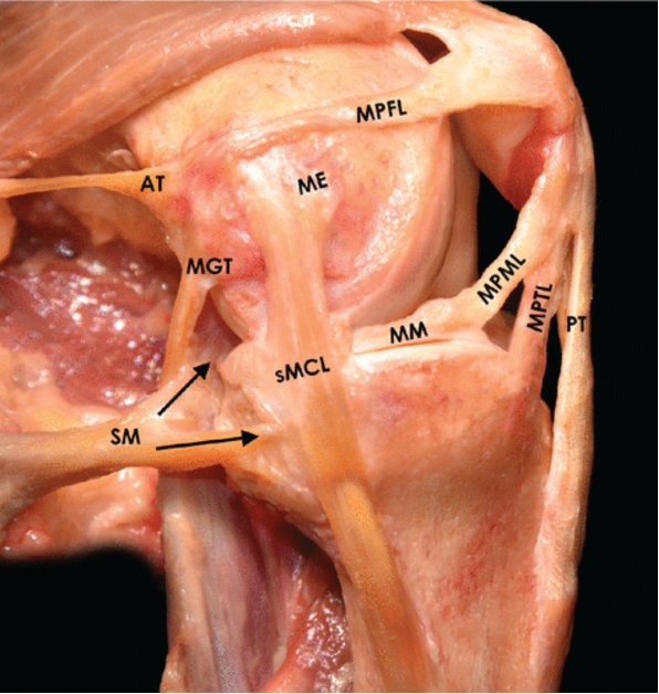
Previous literature is lacking in a description of the medial stabilizing ligaments of the kneecap in relation to surrounding structures and one another. In this study, the attachments of the following ligaments were analyzed in relation to bony and soft tissue structures using cadavers: medial patellofemoral ligament (MPFL), medial patellotibial ligament (MPTL), medial patellomeniscal ligament (MPML), and medial quadriceps tendon femoral ligament (MQTFL). The analysis was performed visually and with x-ray of each specimen. Contrary to what was predicted, each ligament did not have distinct borders. Rather, the MPTL and MPML shared an insertion point on the kneecap, and the MPFL shared an attachment to the underside of one of the quadriceps muscles. The MPTL was also found to attach to the tibia much closer to the joint line than previously described (5mm distal to the joint line vs 10-20 mm). Moreover, this study led to the identification of a bony ridge on the tibia where the MPTL inserted. Through use of this study’s results, surgeons can aim to more accurately restore the normal anatomy of the knee for patients with knee instability due to the MPFL, MPTL, MPML, or MQTFL.

Figure 4. Medial view of a left knee at 90° of flexion demonstrating the attachment sites and orientations of the MPFL, MPTL, and MPML. The relationship of the medial patellar ligament’s attachment sites to other medial knee structures can also be appreciated. The arrows indicate the direct and indirect arms of the semimembranosus. AT, adductor tendon; ME, medial epicondyle; MGT, medial gastrocnemius tendon; MM, medial meniscus; MPFL, medial patellofemoral ligament; MPML, medial patellomeniscal ligament; MPTL, medial patellotibial ligament; PT, patellar tendon; SM, semimembranosus; sMCL, superficial medial collateral ligament.
